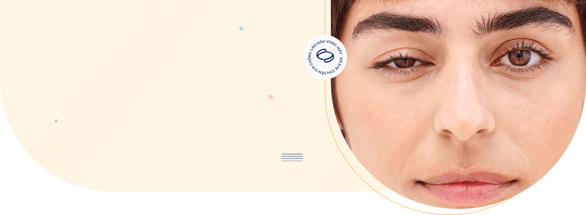
Ptosis, medically termed blepharoptosis, is a structural condition characterized by an unnatural drooping of the upper eyelid. This occurs when the margin of the upper eyelid falls below its normal position, potentially compromising facial symmetry, visual field function, or both.
What is ptosis (Droopy Eyelid)?
Clinically, ptosis is identified when the upper eyelid margin covers the pupil by more than 2 mm, or when the Margin Reflex Distance 1 (MRD1—the clearance between the central corneal light reflex and the upper eyelid edge) measures less than 4 mm.
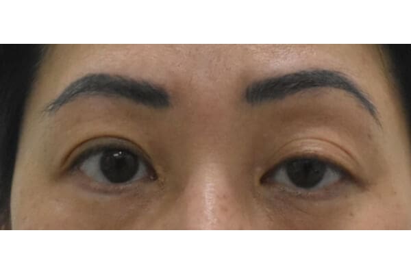
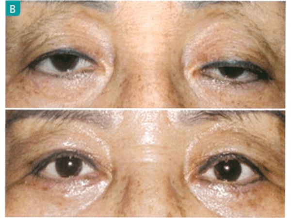
Customer with upper eyelid covering the pupil a lot
The upper eyelid covering 1-2mm of the upper edge of the pupil is normal. When ptosis occurs, the upper eyelid covers the pupil by more than 2 mm, causing poor aesthetics.
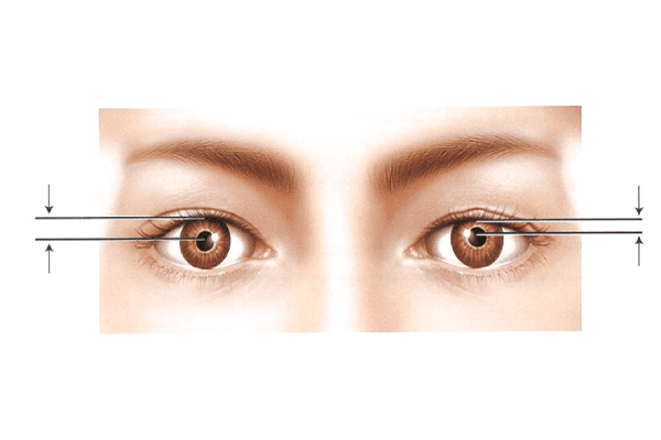
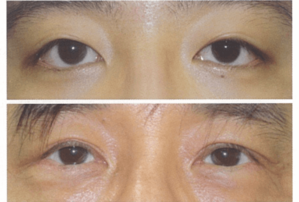
Customer with short MRD1 distance
The distance from the corneal center to the upper eyelid margin MRD1 in a normal eye is from 3-5mm. When the eyelid is drooping, MRD1 <4mm, making the eyes look smaller.
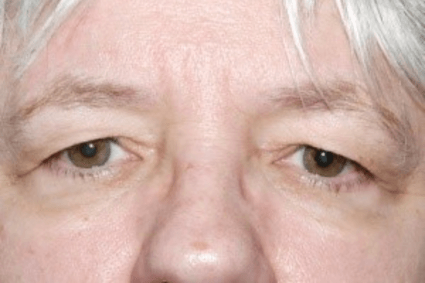
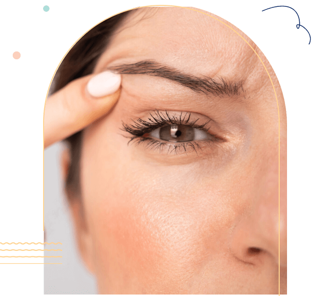
Customer with skin laxity causing ptosis
The skin of the upper eyelid is aging, lax, and sagging down onto the eyeball, causing ptosis. This is a common condition in middle-aged women.
Clinical Classification of Ptosis
Oculoplastic specialists classify blepharoptosis across three distinct metrics to determine the optimal corrective surgical pathway:
– By Degree of Corneal Coverage:
Mild Ptosis: The upper eyelid margin overlaps the pupil by 2 mm or less.
Moderate Ptosis: The upper eyelid covers approximately 3 mm of the pupil.
Severe Ptosis: The upper eyelid covers 4 mm or more of the pupil, significantly obstructing the superior visual field.
– By Upper Eyelid Position (Gaze Assessment):
Grade 1: The eyelid rests slightly below its ideal position, just above the pupil margin (lowered by roughly 2 mm).
Grade 2: The eyelid margin covers the upper portion of the pupil without crossing its center (lowered by 3 mm).
Grade 3: The margin drops past the center of the pupil (lowered by 4 mm or more).
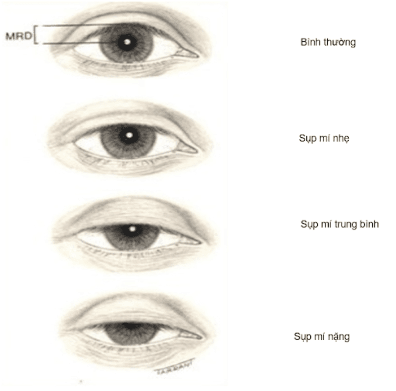
– By Distance from the Corneal Center to the Upper Eyelid Margin (MRD1)
Mild Ptosis: MRD1 measures between 2 mm and 4 mm.
Moderate Ptosis: MRD1 is reduced to a range of 0 mm to 2 mm.
Severe Ptosis: MRD1 is 0 mm or less, indicating the eyelid margin rests at or below the center of the pupil.
Degrees of eyelid ptosis based on the distance from the center of the cornea to the upper eyelid margin MRD.
What Causes Droopy Eyelids (Ptosis)?
Congenital Ptosis
Present from birth, this form stems from developmental deficiencies in the levator palpebrae superioris muscle. It may manifest as isolated myogenic weakness or as part of broader complex presentations like Blepharophimosis, Ptosis, and Epicanthus Inversus Syndrome (BPES).
Acquired Ptosis
Acquired ptosis develops later in life and is more common in adults. It may result from age-related weakening of the levator tendon (aponeurotic ptosis), long-term contact lens use, nerve disorders such as Myasthenia Gravis, eye trauma, or severe upper eyelid skin laxity (dermatochalasis), which can create the appearance of a drooping eyelid (pseudoptosis).
How Does a Droopy Eyelid Impact Your Daily Life?
1
Drooping eyelids on one side affecting eyelid symmetry and overall facial appearance.
2
Drooping upper eyelids narrow your upper field of vision, making daily tasks like driving, reading, and working on screens noticeably fatiguing.
3
Prolonged untreated ptosis in long-term cases can correlate with or exacerbate astigmatism, refractive errors, or strabismus.
4
To compensate for obstructed vision, patients frequently hyperextend their frontalis (forehead) muscles or tilt their heads backward. This chronic overactivation directly causes persistent forehead tension, neck strain, and tension headaches.
Advanced Surgical Treatments for Ptosis Correction
True structural ptosis does not resolve on its own and requires targeted surgical repositioning by an experienced oculoplastic specialist. Depending on your baseline levator muscle function, one of the following evidence-based techniques will be utilized:
Levator Muscle Advancement
This precise technique repairs and tightens the primary lifting muscle of the upper eyelid. It is highly effective for patients who still retain moderate-to-good levator muscle function, restoring a naturally contoured, symmetric double eyelid crease.
However, with this method, droopy eyelids may easily recur or have the potential complications of conjunctival prolapse, eyelid gap, etc. if not performed by a skilled doctor.
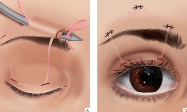
Frontalis Sling / Suspension Procedure
When the native levator muscle is profoundly weak or completely non-functional, the eyelid is mechanically linked to the forehead muscle using specialized biocompatible silicone rods, autologous fascia lata, or banked temporal fascia. This enables the patient to comfortably elevate their eyelids using natural eyebrow movement.
However, if not performed according to standard procedures, this method may carry risks of eyelid gap causing corneal inflammation, pulling the eyelids away from the eyeball, etc.
Direct Frontalis Suspension
The doctor uses the contraction force of the forehead muscle to directly lift the eyelids, applied in cases where the function of the upper eyelid lifting muscle is weak or no longer exists. This method uses autologous material to ensure maximum safety, long-lasting effectiveness even for severe conditions, with less recurrence later.
But this technique is quite complex, requiring a doctor with good expertise, well-versed in the anatomy of the upper eyelid and surrounding structures.
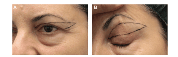
Management of Pseudoptosis (Correcting Excess Eyelid Skin / Dermatochalasis)
When drooping is caused by age-related skin laxity rather than a weak muscle, an upper blepharoplasty or structural brow lift is indicated. This procedure carefully removes redundant skin and fat to unburden the eyelid margin, opening up the eye area cleanly.
Note, patient need to seek reputable facilities for doctors to evaluate, measure, and perform the procedure accurately, ensuring maximum effectiveness and safety.
Why Choose Dr. Eye for Specialized Ptosis Care?
Correcting ptosis requires an intricate balance between structural protection and aesthetic refinement. Our specialized center offers:
Evidence-Based Oculoplastic Consultations
Your care pathway begins with comprehensive assessments of muscle function, corneal sensitivity, and tear film stability.
Tailored Pre-Operative Outlining
Every corrective blueprint is fully customized to your facial proportions, prioritizing functional safety alongside a refreshed, balanced appearance.
Specialized Reconstructive Expertise
Procedures are directed by accredited surgeons possessing extensive experience in delicate ophthalmic and periocular reconstructions.
Individualized Treatment Approaches with a Focus on Safety
Dr. Eye's doctors continuously improve their skills, update the latest technologies, and choose minimal intervention methods, prioritizing your safety above all.
Rigorous Safety and Sterilization Standards
We operate within advanced surgical suites utilizing precise micro-instruments, minimizing trauma and reducing recovery downtimes.
Client Story
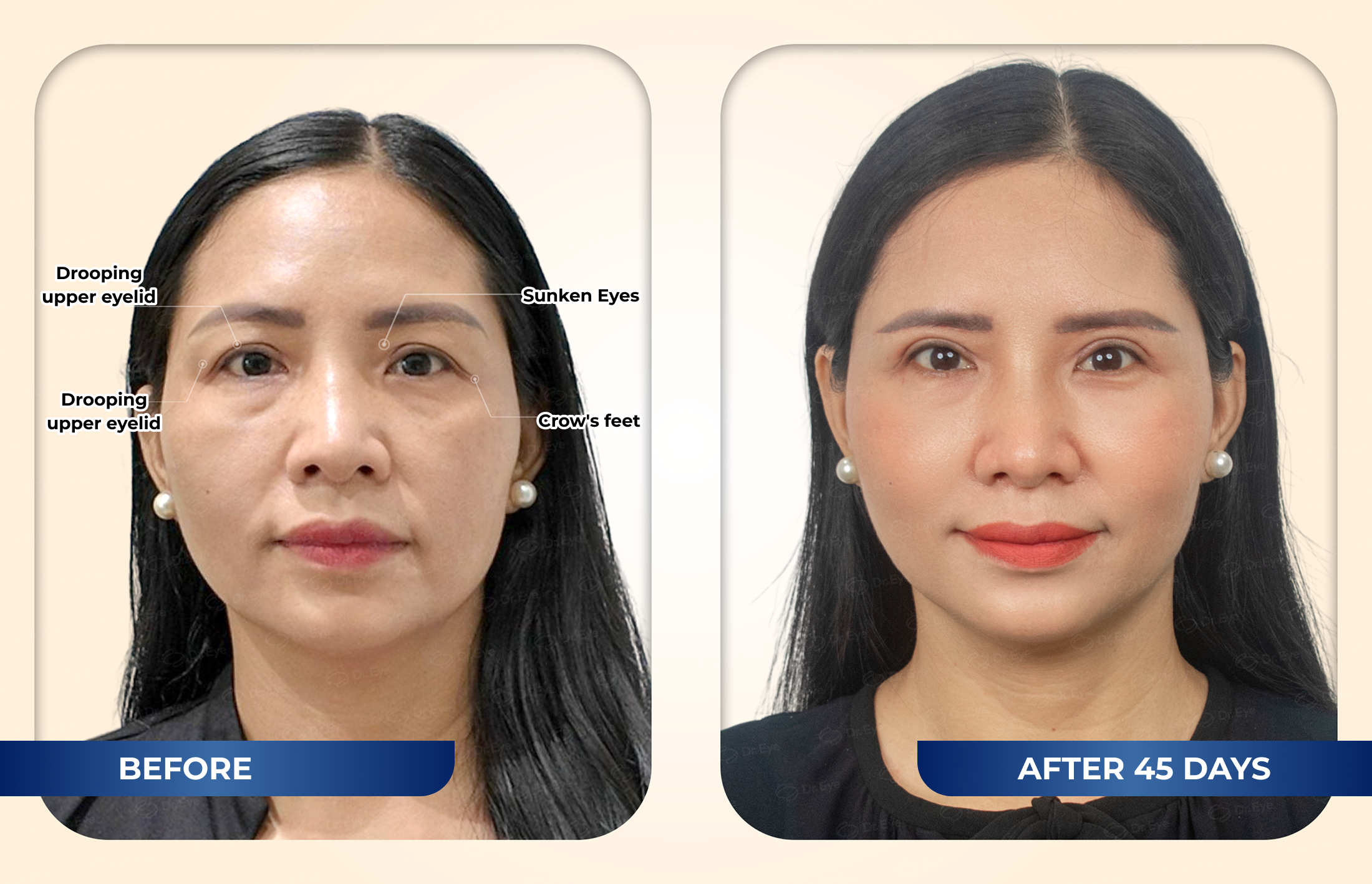
Ms. Dao Phuong Nhi
44 years old, Ho Chi Minh City
EXCESS SKIN REMOVAL ON THE UPPER EYELID – UPPER EYELID FAT GRAFTING – BROW LIFTING UNDER THE BROW ARCH
“I chose Dr. Eye because Dr. Eye specializes in anti-aging treatments for the eye area. When specializing, the doctors have a lot of experience, so I feel assured. Moreover, when I came for a consultation, the doctor provided very accurate, honest, and detailed advice, offering solutions that I found reasonable, so I have confidence. All imperfections were addressed, and I feel my eyes are younger and brighter; looking in the mirror, I feel like I have returned to being 18 years old.”
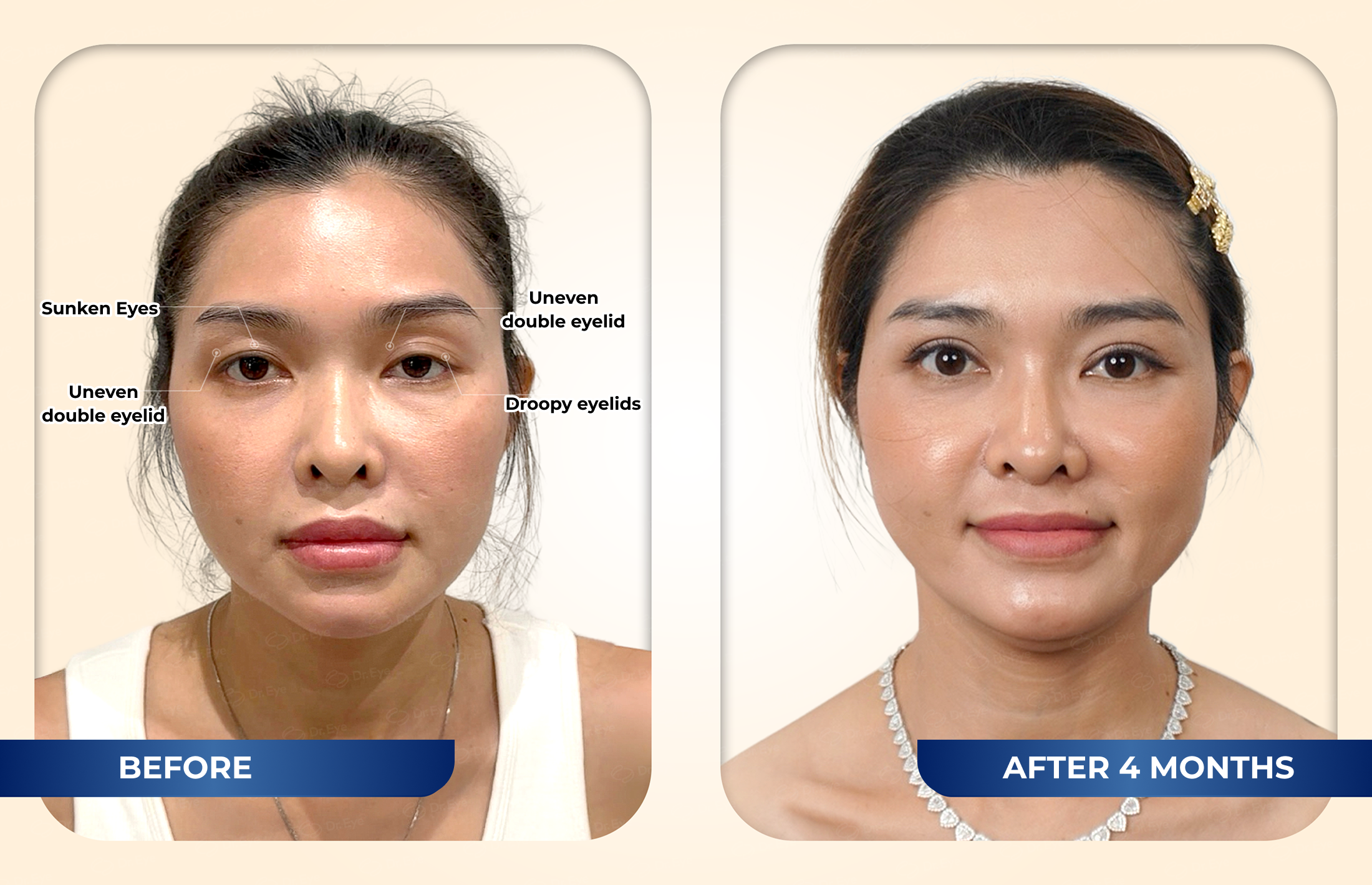
UPPER EYELID SURGERY – TREATMENT FOR DROOPY EYELIDS – UPPER EYELID FAT GRAFTING
“I had previously made a deposit elsewhere but did not proceed because I felt their consultation was superficial. However, when I came to Dr. Eye for a consultation, I felt that the doctor had high expertise, explained everything in detail, and advised me on what to do and what not to do. I felt reassured and decided to proceed at Dr. Eye. The results after 1 month are very satisfactory; I find it beautiful, natural, and my eyes look more soulful, I feel confident and more energetic.”
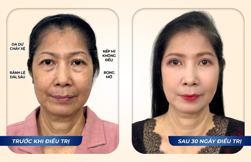
LIFTING EYEBROWS – PTOSIS TREATMENT – LOWER EYELID SURGERY
“I have wanted to do my eyes for a long time, not just recently, but I haven’t had the trust, and coming to Dr. Eye has been the 7th or 8th place already. Then, by chance, as if it were fate, I saw Dr. Eye’s Facebook page specializing in aging eyes, aimed at middle-aged people like my age, so I researched, scheduled a day to consult, and then decided to do cosmetic surgery right away.
Understanding the desire to preserve the youthful beauty of the eyes for middle-aged women, Dr. Eye offers modern solutions to address all aging issues around the eyes such as sagging eyelids, eye bags, crow’s feet,…
Care and Preservation of Upper Eyelid Health
While structural muscle weakness requires surgical repair, maintaining healthy periocular tissue can streamline your recovery and protect overall tissue elasticity:
1. Prioritize Consistent Rest: Ensure adequate sleep cycles to reduce chronic vascular strain and fluid retention around the eyes.
2. Incorporate Screen Breaks: Follow the 20-20-20 rule during prolonged digital device use to facilitate regular blinking and mitigate dry eye strain.
3. Practice Gentle Eye Care: Avoid aggressive rubbing of the eyelids, which can prematurely stretch the fragile levator aponeurosis over time.
4. Protect Against UV Damage: Apply a broad-spectrum sunscreen daily and wear UV-protected sunglasses when outdoors to shield delicate skin from premature breakdown.
5. Increase the intake of foods rich in vitamin A, vitamin E, and other nutrients beneficial for eye health.
The above information addresses the condition of droopy eyelids. If you are experiencing a similar situation, don’t hesitate to visit Dr. Eye for our specialized team of doctors to provide appropriate remedies, helping to restore both eyelid function and natural ocular symmetry.


 BOOK A SPECIALIST CONSULTATION